Figure 6.
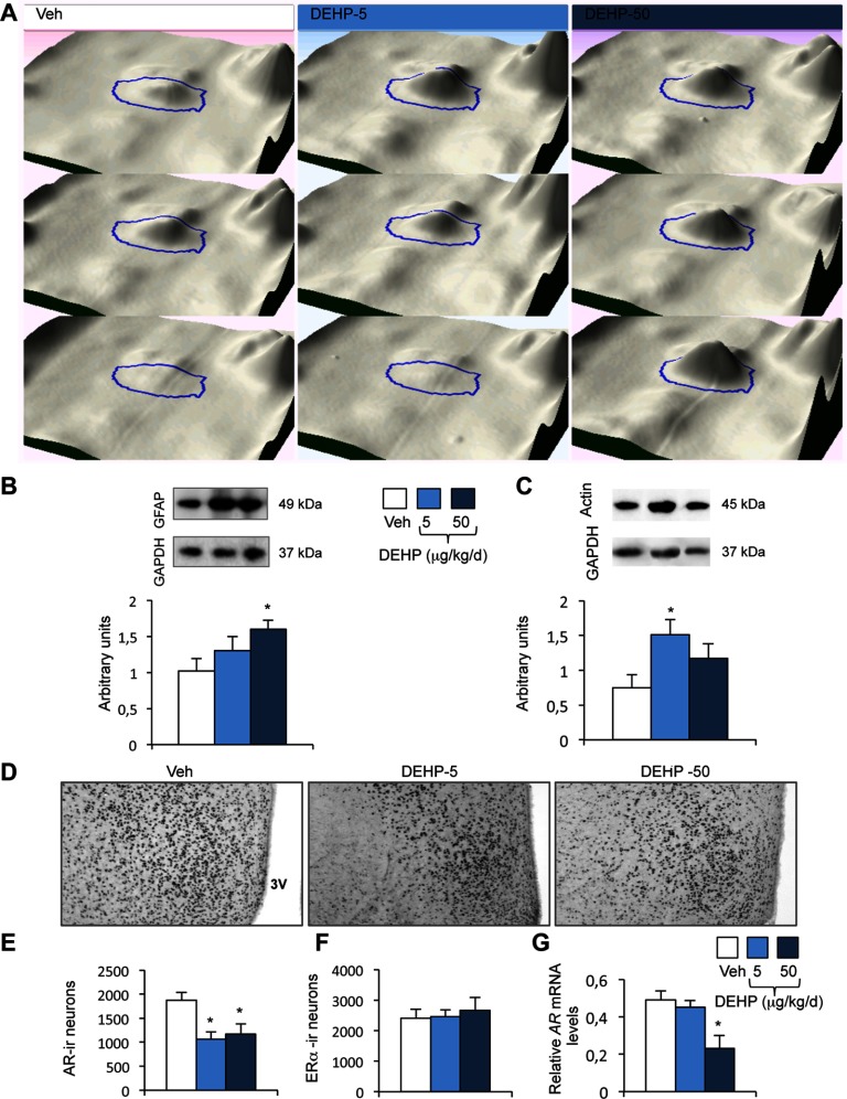
Validation of proteomic data and characterization of androgen receptor (AR) and expression in the medial preoptic nucleus (MPN). (A) 3-D view of the spot ID:0656, corresponding to GFAP, shown for three animals exposed to the vehicle (Veh) or DEHP at 5 or . (B–C) Upper panels: Representative Western blots of GFAP (B), (C), and GAPDH used as a protein reference, in the MPN of the Veh, DEHP-5, and DEHP-50 groups. Lower panels: quantification of the protein levels normalized to GAPDH. Data are expressed as the means of four males per treatment group, * compared to the vehicle group. (D–F) Representative AR-immunolabeling of the medial preoptic nucleus of males exposed to the vehicle (Veh) and DEHP at 5 or (D) and quantitative data for AR- (E) and cells (F). 3V: Third ventricle. Data are expressed as the means of four to six males per treatment group, * vs. the vehicle group. (G) AR gene expression in males exposed to the vehicle and DEHP at 5 or . Data are expressed as the means of six to eight males per treatment group, * compared to the vehicle group.

