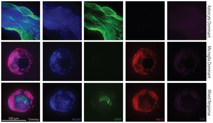
FIG. 4.
Immunohistochemical images of individual CSF intake holes from 3 different explanted ventricular catheters demonstrating representative astrocyte-dominant, microglia-dominant, and mixed responses. All images obtained with 10× objective, 499.2-μm z-stack, 2.4-μm step size. Figure is available in color online only.

