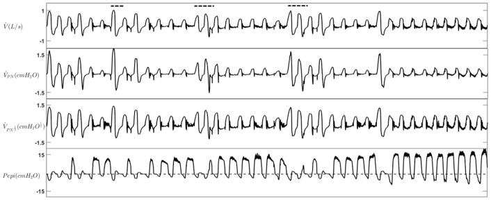
Figure 5.
An example tracing of expiratory flow limitation during NREM sleep. Flow was measured with both a pneumotachograph (V̇) and nasal cannula (V̇PN). Nasal pressure was also linearized using the square root transform ( ). During EFL periods, expiratory positive airway pressure developed at the level of the epiglottis (Pepi) without any increase in expiratory flow. Dashed lines show the scored arousals.

