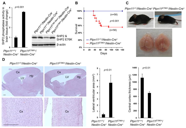
Fig. 1. Neural cell–specific expression of Ptpn11E76K induces hydrocephalus and brain developmental defects.
(A) Brain tissues dissected from one-month-old mice (n=3 mice per genotype) were lysed, and SHP2 catalytic activities in the lysates were assessed by the immunocomplex phosphatase assay. SHP2 abundance in the lysates was examined by immunoblotting. (B) Kaplan-Meier survival curves of mice of the indicated genotypes. (C) Representative images of one-month-old mice and their brains. (D) Sagittal brain sections were processed for H&E staining. Analyses in all panels were performed in 3 independent experiments, n=4 mice per genotype, and representative images are shown. Data are presented as mean±S.D. of biological replicates. Cx, cortex; Hp, hippocampus; LV, lateral ventricles. Scale bars, 2 cm (C, mice), 5 mm (C, brain), and 500 μm (D).

