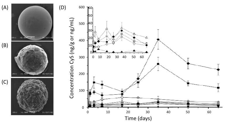
Figure 12.
Scanning electron microscopy images (SEM) of PLGA microparticles loaded with cyclosporine (A) SEM image of microparticles before the in vitro release assay (B) SEM image after two weeks of in vitro release (C) SEM image taken after two months of in vitro release (10 μm scale bar). (D) Diagram showing the concentration of cyclosporine released overtime in different ocular tissues and blood subsequently after the cyclosporine microparticles are intravitreally injected for the treatment of uveitis. (×) Iris-ciliary body; (□) cornea; (△) conjunctiva; (○) aqueous humor; (◇) blood; (□) vitreous body; (●) choroid-retina; (◆) sclera; (▲) lens. Reproduced with permission.[86]Copyright 2006, Association for Research in Vision and Ophthalmology.

