Figure 1.
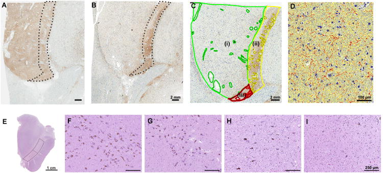
Image analysis of TH-ir in subregions of putamen using ImageScope and semi-quantitative assessment of substantia nigra pigmented neurons. (A) and (B) show tyrosine hydroxylase immunohistochemistry of the putamen at the level of the anterior commissure. In a representative advanced LBD case (B) marked reduction of TH-ir is observed in dorsolateral putamen compared with a neurologically normal control (A). Note that TH-ir in the ventromedial putamen (dashed circle area) is preserved even in advanced LBD. (C): Green-circled area represents dorsolateral putamen (i). Ventrolateral putamen is the combined area of yellow-circled area (medial, (ii)) and red-circled area (ventral, (iii)). For image analysis, artifacts, large fiber tracts and blood vessels were manually edited (dashed circle) on each section. (D) A color deconvolution-based algorithm was used to measure TH-ir. A positive pixel count algorithm was customized to quantify brown immunoreactive pixels (red markup), subtracting inverse (blue) and background pixels (yellow). Transverse section of the unilateral midbrain (E). Warped rectangles show the lateral and medial substantia nigra. Semi-quantitative assessment of neuronal loss in the substantia nigra. Abundant pigmented neurons are seen in a neurologically normal control (Score 0) (F), compared with variable neuronal loss in LBD - mild (Score 1) (G), moderate (Score 2) (H) and severe (Score 3) (I). Scale bars: A - C = 2 mm; D = 100 μm; E = 1 cm; F – I = 250 μm.

