
Figure 1. Spectrum of structural divergence which preserves biochemical function.
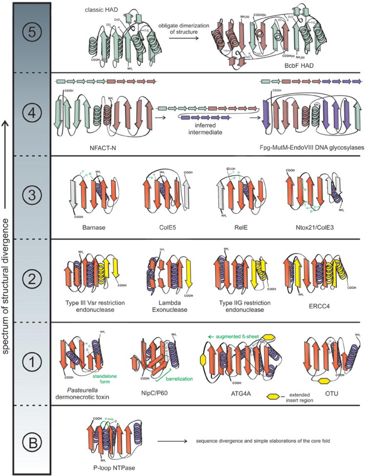
The spectrum is broken up into distinct classes of structural divergence separated by dotted lines. Example structures are depicted as topology diagrams with arrows representing β-strands and coils representing α-helices. ‘B’ represents the ‘baseline’ class of structural divergence. Class 1: representatives of papain-like peptidases; Class 2: restriction endonuclease fold with modified part of the secondary structural elements colored in yellow; Class 3: BECR fold members with divergent active site residues highlighted in green; Class 4: transition between NFACT and Fpg-MutM-EndoVIII DNA glycosylase proteins are accompanied by linear arrays of secondary structural elements to show rewiring, each duplicated basic 4-stranded element is given a distinct color; Class 5: topological transmogrification observed in the obligate dimer-forming BcbF family of HAD domains, strands are labeled, and monomers are given distinct colors.