
Figure 3. Cartoon representations of various STY kinase domains illustrating the structural transformations in the superfamily.
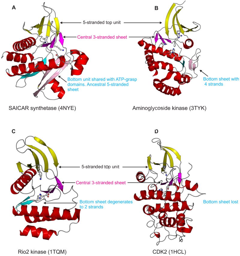
Helices are colored red, whereas strands are colored based on their structural unit. Strands of the top unit are colored yellow and those in the central sheet, magenta. The cyan and pink strands of the bottom unit show the equivalence of these strands between the structures.