Figure 3.
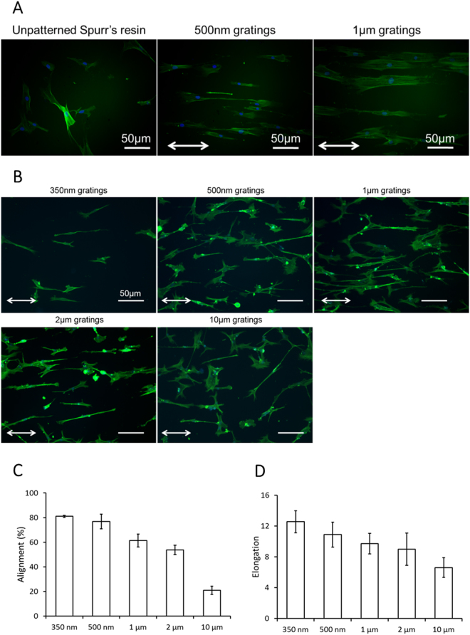
F-actin cytoskeleton visualized by Oregon-green labeled phallodin with DAPI as counter-stain for cell nuclei in hMSC on (A) unpatterned Spurr’s resin, Spurr’s resin with 350 nm gratings and 500 nm gratings, (B) PDMS with 350 nm, 500 nm gratings, 1 µm, 2 µm and 10 µm gratings. (Bar = 50 μm. Images are taken with fluorescence microscopy. White arrows denote the grating axis direction.) Effect of groove grating width on hMSC morphological properties. Changes in cell body (C) alignment and (D) elongation of hMSCs cultured on PDMS gratings with different groove widths. (Bars denote standard deviation).

