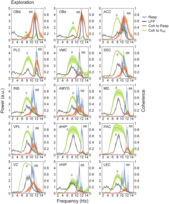
Figure 5.
Parallel detection of theta (θ) and respiration-entrained LFP rhythm (RR) throughout the mouse brain during exploration. Panels show the same as in Fig. 2. Each sample consisted of 30 seconds of concatenated data obtained during exploration with respiration faster than theta. The reference theta-filtered signal was taken from either the dorsal hippocampus or the parietal cortex. Respiration was assessed through thermocouples in the nasal cavity.

