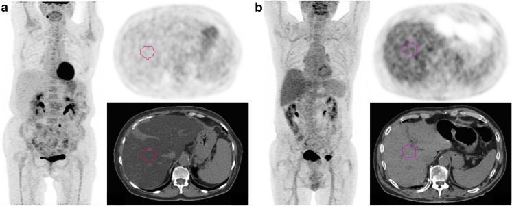
Fig. 4.
Representative examples of steatotic (a) and non-steatotic (b) patients. Maximum intensity projection (MIP) and trans-hepatic axial PET and CT images of 67 year-old female steatotic patient addressed for an interim PET of DLBCL scored DS5 (a) and a 84-year-old male non-steatotic patient addressed for EoT PET of a DLBCL scored DS1 (b). The automatic 3 cm-diameter VOIs in the right liver lobe are displayed in pink. Images are scaled on the same maximum value. Note that the intense FDG focus on the left groin of the patient illustrated on panel b is a benign uptake due to a plug

