FIGURE 4.
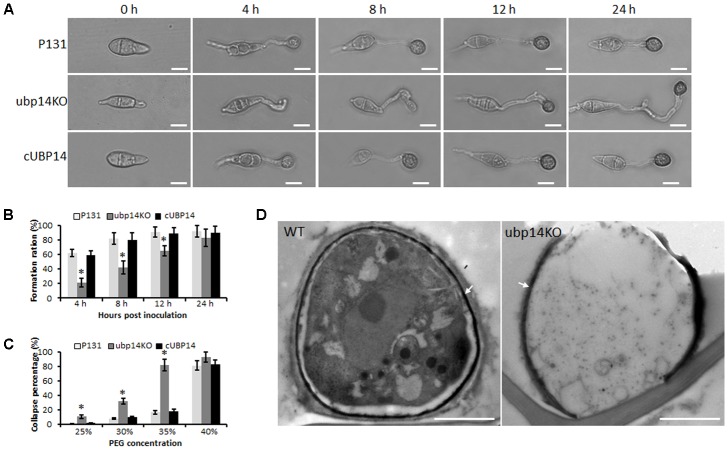
MoUBP14 affects functions of appressorium. (A) Appressorium formation assay. Conidia were incubated on hydrophobicsurfaces and observed at different time points. Bar, 10 μm. (B) Appressorium formation rates at different time. Asterisks representsignificant differences (P < 0.05, n > 100). (C) Cytorrhysis assay for appressorium turgor pressure. Drops of conidial suspension (1 × 10⁵ conidia ml-1) were placed on the hydrophobic surface of coverslips and treated with indicated concentration of PEG8000 at 24 hpi. (D) Transmission electron microscopy (TEM) observation of the appressorium. Melanin layer was indicated by arrows.

