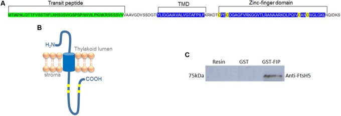
FIGURE 1.
FIP is a transmembrane protein containing a zinc-finger domain and is localized in the thylakoid membrane. (A) Amino acid sequence of FIP protein. The transit peptide, transmembrane domain (TMD), and zinc-finger domain are marked. (B) Theoretical model for FIP topology predicted by PROTTER online software. FIP is anchored in thylakoids by TMD and has an amino proximal luminal domain and a carboxyl proximal stromal domain. (C) glutathione S-transferase (GST) pull-down assay of the FtsH5 and FIP interaction. GST and GST-FIP fusion proteins were expressed in Escherichia coli and purified with a GST column. GST and GST-FIP were incubated with nickel resin-purified recombinant FtsH5 fusion protein, pulled down with GST-beads, and detected using an anti-FtsH5 antibody by Western blot analysis.

