
Figure 2. Bioactivities of PKUMDL-WQ-2101 and PKUMDL-WQ-2201 in cell based assays.
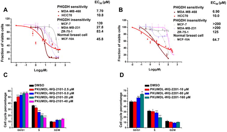
(A-B) Growth inhibition activity of PKUMDL-WQ-2101 (A) and PKUMDL-WQ-2201 (B) in MDA-MB-468, HCC70, MCF-7, MDA-MB-231, ZR-75-1 and MCF-10A cells, respectively. Cells were exposed to vehcile or various concentrations of PKUMDL-WQ-2101 for 72h followed by MTT assay. The EC50 value of PKUMDL-WQ-2201 for MCF-7 and MDA-MB-231 was larger than 200 μM, so the corresponding dose-response curve was not presented here. (C-D) Percentage of MDA-MB-468 cells in different phases of the cell cycle after respectively treatment with 2.5, 5.0, 20 and 40 μM PKUMDL-WQ-2101 (C), and 10, 80 and 160 μM PKUMDL-WQ-2201 (D) for 24 hours. DMSO was used as vehcile. Data represent the mean ± SD independent experiments. Difference is significant by two-tailed multiple t-test, *p < 0.05, **p < 0.01, ***p < 0.001. See Figure S3 for cell bioactivities of PKUMDL-WQ-2202 and 2203.