
Figure 3. CRISPR-Cas9 mediated PHGDH KO and PHGDH inhibition by PKUMDL-WQ-2101 and PKUMDL-WQ-2201.
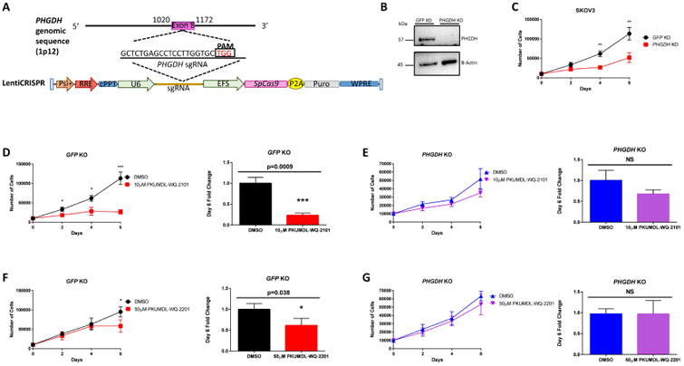
(A)Overview of LentiCRISPR system and sgRNA design generated for targeted PHGDH deletion in SKOV3 ovarian cancer cells. (B) Western blot analysis for SKOV3 GFP KO control and SKOV3 PHGDH KO cells with actin as a loading control. (C) Growth curve comparing SKOV3 GFP KO control and PHGDH KO over 6 days. (D) Growth curves of SKOV3 GFP KO control and (E) PHGDH KO cells after 6 days of treatment with vehicle or 10μM PKUMDL-WQ-2101 followed by cell counting. (F) Growth curves of SKOV3 GFP KO control or (G) PHGDH KO cells after 6 days of treatment with vehicle or 50μM PKUMDL-WQ-2201 followed by cell counting. All values represent the mean ± SEM from n=3 biological replicates. P values were obtained from a two-tailed student's t-test, *P< 0.05, **p<0.01, ***P<0.001. See Figure S4 for PKUMDL-WQ-2101 and 2201 bioactivies on SKOV3 GFP KO cells and results of PKUMDL-WQ-2101 pull down assays.