
Figure 4. PKUMDL-WQ-2101 and PKUMDL-WQ-2201 inhibit the serine biosynthesis pathway in cells.
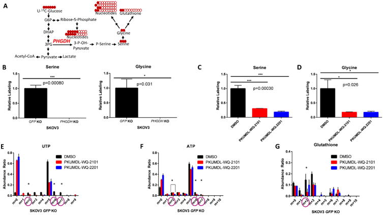
(A) Schematic of U-13C-glucose stable isotope labeling used to detect carbon labeling from glucose (red) in metabolites part of the serine metabolic network. (B) 13C-serine and 13C-glycine labeling from glucose in SKOV3 GFP KO control cells compared to SKOV3 PHGDH KO cells after 24 hours. (C)13C-serine and (D) 13C-glycine labeling from glucose in SKOV3 GFP KO cells after 24 hour treatment with 37 μM PKUMDL-WQ-2101 and 291 μM PKUMDL-WQ-2201, followed by subsequent U-13C-glucose labeling. (E) Mass isotopomer distribution (MID) of UTP and (F) ATP after 24 hour treatment with 37 μM PKUMDL-WQ-2101 and 291 μM PKUMDL-WQ-2201, followed by subsequent U-13C-glucose labeling. (G) Glutathione after 24 hour treatment with 37 μM PKUMDL-WQ-2101 and 291 μM PKUMDL-WQ-2201, followed by subsequent U-13C-glucose labeling. All values represent the mean ± SEM from n=3 biological replicates. Difference is significant by One-Way ANOVA, *P< 0.05, **p<0.01, ***P<0.001.