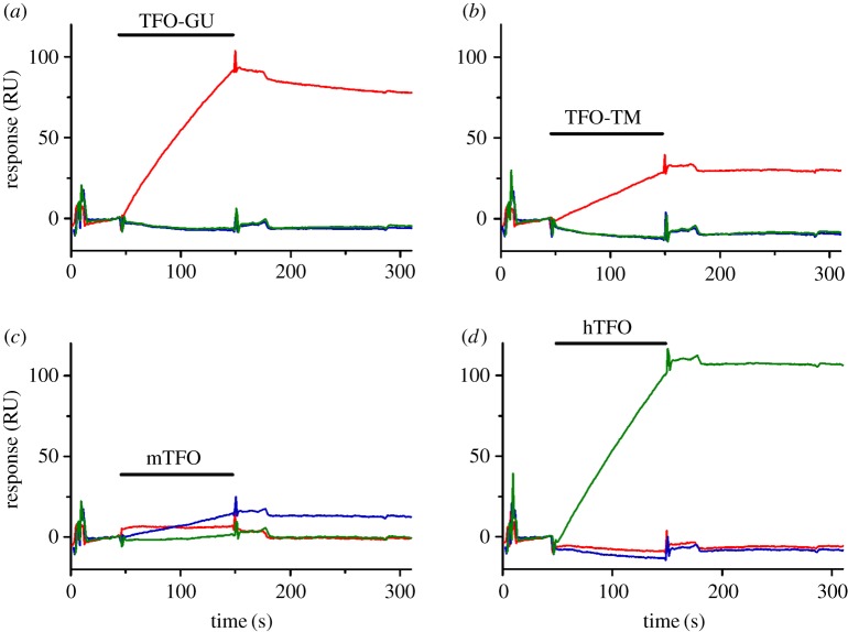
Figure 2.
Surface plasmon resonance (SPR analysis) of TFO binding to immobilized DNA targets at 37°C in MES buffer, pH 6.5. The sensorgrams show complex formation following injection of 35 ml 4 mM TFO (indicated by a black horizontal bar; (a) TFO-GU (targeting VEGF); (b) TFO-TM (targeting VEGF); (c) mTFO (targeting mGAA); (d) hTFO (targeting human GAA (hGAA)) over surfaces containing immobilized biotinylated hGAA (green line), mGAA (blue line) and VEGF (red line) target hairpin duplexes, after which dissociation of the complexes is monitored [41].

