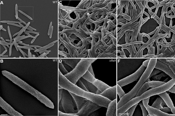
FIG 3 .
Altered cell surface of the ftsX and envC mutants revealed by scanning electron microscopy. (A to F) Exponential-phase F. nucleatum cells. Wild-type (A and B), ΔftsX (C and D), and ΔenvC (E and F) strains were immobilized on coverslips and fixed with 2.5% glutaraldehyde prior to viewing by a scanning electron microscope. Enlarged areas of panels A, C, and E are shown in panels B, D, and F, respectively. Bars, 1 µm (A, C, and E) and 0.5 µm (B, D, and F).

