
Figure 4.
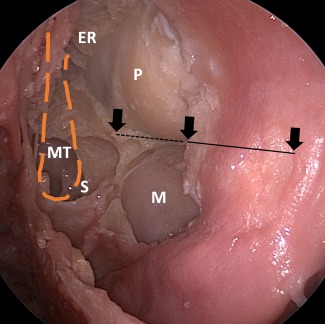
Endoscopic view of the left orbit following modified inferomedial orbital strut technique (IOS) (cadaver). The arrow represents the anterior, middle and posterior aspect of the IOS. The dotted line is the posterior half of the IOS that has been removed. ER = ethmoid roof; M = maxillary sinus; MT = previously resected area of the middle turbinate; P = periorbita; S = sphenoid sinus.