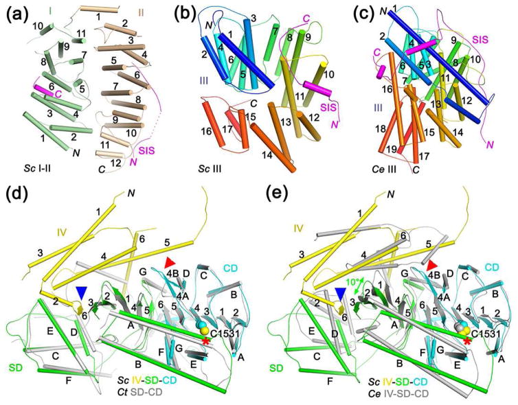
Figure 2.
Structures of separase domains and their interaction with securin. (a). Schematic drawing of domains I and II of yeast separase. The segment of SIS that interacts with these domains are also shown (magenta). (b). Schematic drawing of domain III of yeast separase, colored from blue at the N-terminus to red at the C-terminus. (c). Schematic drawing of domain III of C. elegans separase. (d). Overlay of the structures of yeast IV-SD-CD (in color) and the SD-CD of C. thermophilum separase (gray). The catalytic Cys residue is indicated with the red asterisk. Blue arrowhead points to the additional β-strand provided by domain IV to the SD in yeast separase. Red arrowhead points to conformational differences in the loop L4 region. (e). Overlay of the structures of yeast IV-SD-CD (in color) and the IV-SD-CD of C. elegans separase (gray). The superposition is based on the CD only, and a 10° difference is observed for the forientation of the SD β sheet (green arrow).

