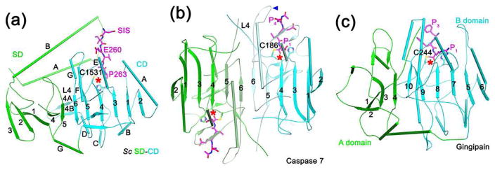
Figure 4.
Structural comparisons with caspase and gingipain. (a). Schematic drawing of SD-CD of yeast separase (green and cyan, respectively), together with the portion of securin SIS (magenta) that is in the CD active site (indicated with the red star). The four-helical bundle in SD is omitted for clarity. (b). Schematic drawing of caspase 7 in a covalent complex with a substrate-mimic inhibitor (magenta). One molecule is colored in light cyan and cyan for its two fragments, while the other in light green and green. The blue arrowhead indicates the loop preceding strand β6 that is in the active site. The orientation of the first molecule is the same as that of separase CD. (c). Schematic drawing of gingipain in a covalent complex with a substrate-mimic inhibitor (magenta). The A and B domains are colored in green and cyan, respectively.

