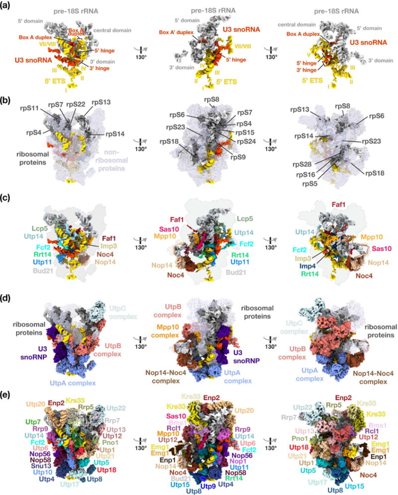
Figure 2.

Structural organization of the yeast small subunit processome. (a) RNA molecules of the SSU processome are shown as surfaces with 5′ ETS (yellow), U3 snoRNA (red) and pre-18S (light-grey). Structural elements of RNAs and helices of the 5′ ETS are indicated. (b) Ribosomal proteins are represented in dark-grey, non-ribosomal assembly factors in transparent light-blue, and RNA species as in (a). (c) Surface representation of centrally located ribosome assembly factors. (d) Visualization of the complexes UtpA (blue), UtpB (red), U3 snoRNP (purple), UtpC (light-blue), the Nop14-Noc4 complex (brown) and the Mpp10 complex (orange). (e) Surface representation of all individual components of the small subunit processome.
