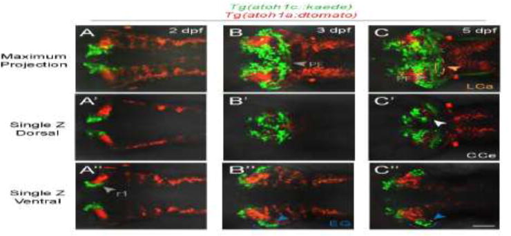
Figure 4. atoh1a- and atoh1c-derived neurons specify distinct progenitor pools.

Live imaging of wild-type embryos with Tg(atoh1c∷kaede) (green) and Tg(atoh1a:dtomato) (red) transgenes indicate atoh1a and atoh1c-derived neurons are distinct cerebellar populations. Maximum projections of a z-stack (A–C) and single z-slices at dorsal (A′–C′) or ventral (A″–C″) focal planes with anterior to the left. A: 2 dpf, B: 3 dpf, C: 5 dpf. Scale bar: 50 μM.
