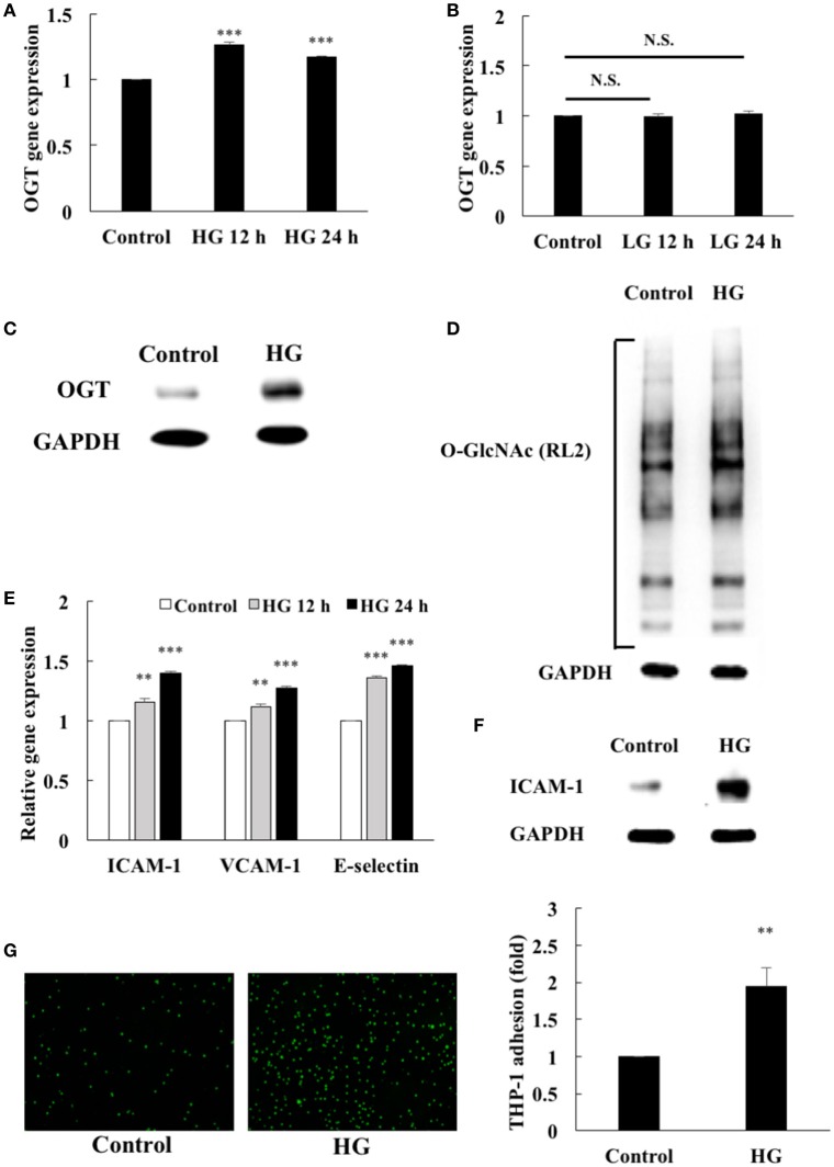
Figure 1.
Endothelial OGT expression, protein O-GlcNAcylation, and inflammatory phenotypes were induced by high glucose (HG). (A) HAECs were stimulated with HG (25 mM) for 12 and 24 h. Real-time PCR revealed that HG stimulation induced 1.27- and 1.17-fold increases, respectively, in OGT mRNA levels N = 5. ***p < 0.001 compared with control. (B) L-Glucose (LG) (25 mM) did not modulate OGT mRNA levels N = 4. N.S. not significant. (C) Stimulation of the HAECs with HG for 24 h induced a significant increase in OGT expression. The blot is representative of three independent experiments. (D) Stimulation of the HAECs with HG for 24 h induced a significant increase in protein O-GlcNAcylation as detected using the RL2 antibody. The blot is representative of three independent experiments. (E) Stimulation of the HAECs with HG for 12 and 24 h induced 1.15- and 1.40-fold increases, 1.11- and 1.27-fold increases, and 1.36- and 1.46-fold increases in ICAM-1, VCAM-1, and E-selectin gene expression levels, respectively N = 5. **p < 0.01 and ***p < 0.001 compared with control. (F) Stimulation of the HAECs with HG for 24 h induced a significant increase in ICAM-1 expression. The blot is representative of three independent experiments. (G) Stimulation of the HAECs with HG for 24 h induced a 1.95-fold increase in THP-1 adhesion to HAECs N = 4. **p < 0.01 compared with control.

