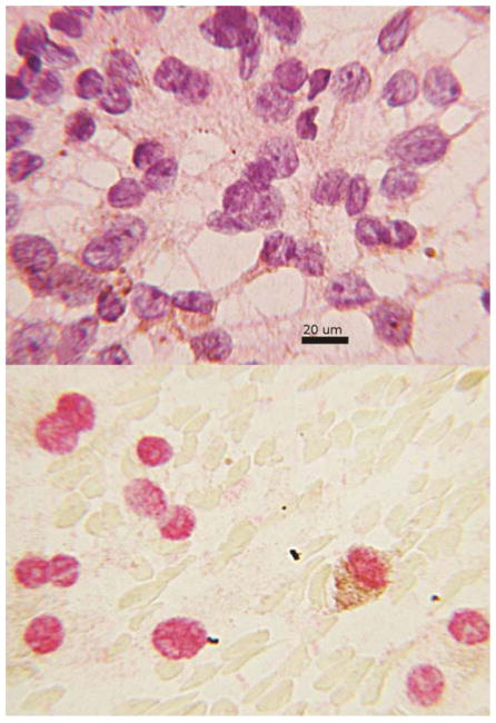
Figure 2.
Figure 2A., above cytology smear from a fine needle aspirate diagnosed as melanoma. Enlarged nuclei, prominent nucleoli and brown cytoplasmic pigment are predominant features. The cytoplasm tapers in a dendritiform pattern, hematoxylin and eosin. B., below, same case reacted with anti-BAP1 by immunocytochemistry. The red staining areas indicate the presence of protein is localized to the nucleus. The melanin pigment appears brown and features the same dendritiform configuration seen in 2A.

