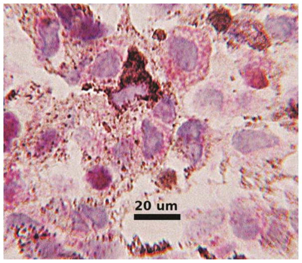
Figure 3.
Immunocytochemical reactivity for anti-BAP1 from a fine needle aspirate of uveal melanoma shows red cytoplasmic granules. In some cells the red color appears to overlie the nucleus and obfuscating the interpretation. Nuclei did not appear to contain red chromogen in cells without accompanying cytoplasmic reactivity suggesting interpretation as a negative reaction.

