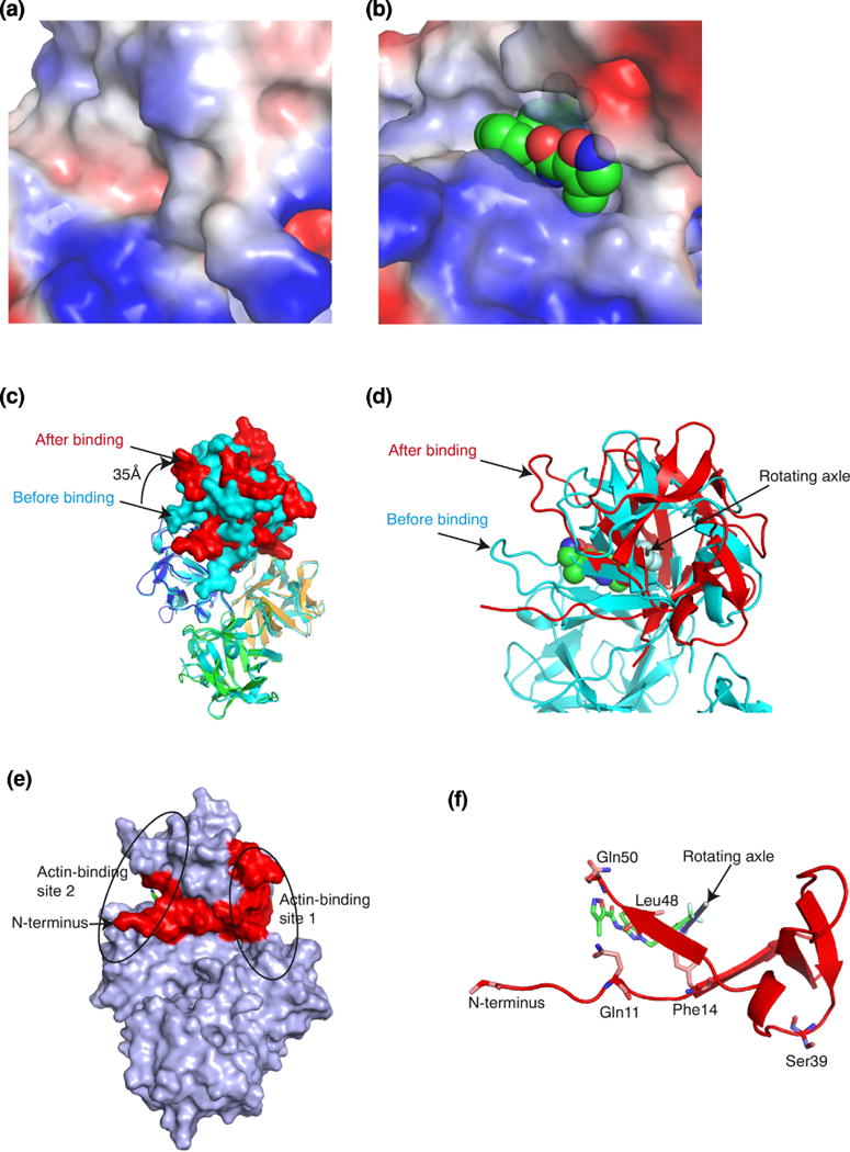
Figure 4.

NP-G2-029 induced changes in fascin conformation. (a) Structure of the actin-binding site 2 in the absence of NP-G2-029. (b) Structure of the actin-binding site 2 with bound NP-G2-029. (c) Superposition of fascin structures in the absence or presence of NP-G2-029. The color marking of the 4 domains of fascin in the presence of NP-G2-029 is the same as in Fig. 2c. The structure of fascin in the absence of NP-G2-029 is colored in light blue. Relative to the location in the absence of NP-G2-029, domain 1 rotated ~35Å clockwise in the presence of NP-G2-029. (d) The rotating axle of domain 1 is marked by a rod. (e) The N-terminal (marked in red) of fascin couples the actin-binding sites 1 and 2. (f) The N-terminal of fascin participates in the binding of NP-G2-029.
