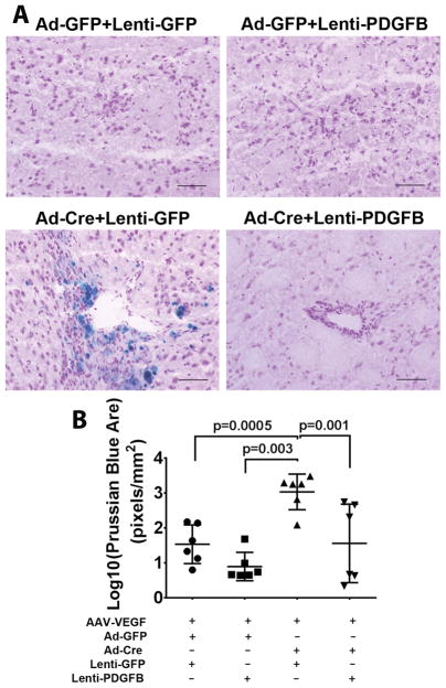
Figure 6. Overexpression of PDGFB reduced hemorrhage in the bAVM lesions.
A. Representative images of Prussian blue stained sections. Scale bar: 100 μm. B. Quantification of Prussian blue positive area. The data are 10 log conversed (Log 10). All mice are treated with AAV-VEGF to induce brain angiogenesis. Ad-GFP+Lenti-GFP: Controls for normal angiogenesis; Ad-GFP+Lenti-PDGFB: Overexpressing PDGFB in angiogenic brain; Ad-Cre+Lenti-GFP: Untreated bAVM; Ad-Cre+Lenti-PDGFB; Overexpression of PDGFB in bAVM. N=6. P values were determined by one-way ANOVA followed by Sidak’s multiple comparisons.

