
Figure 2.
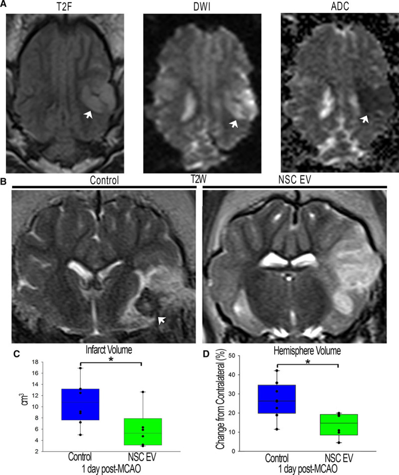
Neural stem cell–derived extracellular vesicle (NSC EV) treatment decreases intracranial hemorrhage, lesion volume, and hemispheric swelling 1-day post-middle cerebral artery occlusion (MCAO). T2-weighted (T2W) and diffusion-weighted imaging (DWI) sequences revealed territorial hyperintense lesions characteristic of an edematous injury (A, white arrows). Hypointense lesions observed on corresponding apparent diffusion coefficient (ADC) maps confirmed areas of restricted diffusion indicative of cytotoxic edema (A, white arrow). These resulting hallmarks demonstrated permanent cauterization of the ventral aspect of the middle cerebral artery resulted in bona fide, repeatable ischemic stroke in all pigs. NSC EV–treated pigs exhibited a reduced incidence of intracranial hemorrhage (B, white arrows). NSC EV–treated pigs also demonstrated a significant (P<0.05) decrease in edema-corrected lesion volume when compared with control pigs at 1-day post-MCAO (6.0±1.4 vs 10.7±1.4 cm3, respectively; C) and a significantly (P<0.01) lower percent increase in hemisphere volume resulting in a less pronounced midline shift relative to control pigs at 1-day post-MCAO (113.77%±2.571% vs 126.83%±3.41% respectively; D). *Significant difference between treatment groups.