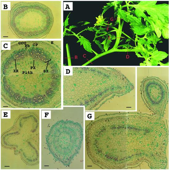
Figure 4.
Anatomical analysis of an epiphyllus inflorescence in clau mutant. A, An epiphyllus inflorescence. Red bars B through G correspond to sites of cross sections shown in B through G. B, A cross-section through a stem showing ring-shaped vascular tissues. Bar = 560 μm. C, A cross-section through a petiole-like organ showing ring-shaped vascular tissues characteristic of a stem. Bar = 220 μm. D, A cross-section through a rachis showing U-shaped vascular tissues. Bar = 220 μm. E, A cross-section through a shoot-like structure emerging from a rachis demonstrating fusion of three petiolules, each with U-shaped vascular tissues. Bar = 560 μm. F, A cross-section through a flower-carrying stem (pedicle). Bar = 130 μm. G, A cross-section through the fused structure of a petiolule (U-shaped) and the inflorescence stem (peduncle; ring-shaped). Bar = 220 μm. CCO, Cortex collenchyma; CP, cortex parenchyma; E, epidermis; Ph, phloem; PX, primary xylem; SX, secondary xylem; XR, xylem rays.

