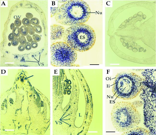
Figure 7.
In situ localization of LeT6 RNA in wild-type (A–C) and clau mutant (D–F) carpels. A, A longitudinal section of a wild-type carpel at anthesis showing expression of LeT6 in vascular tissues and in a distinct region of the ovule integument. Bar = 250 μm. B, A higher magnification of wild-type ovules at anthesis showing the confinement of LeT6 RNA to the inner part of the integument. Bar = 50 μm. C, A longitudinal section of a wild-type carpel post-anthesis. Bar = 400 μm. D, A longitudinal section of a clau mutant carpel post-anthesis showing LeT6 RNA in ovules and vascular tissues. Arrow indicates an ectopic meristem near the stylar end. Note the typical multiloculed ovary arranged in tiers. Bar = 400 μm. E, A higher magnification of the mutant ovary wall showing expression in vascular tissues. Bar = 250 μm. F, A higher magnification of mutant ovules post-anthesis showing LeT6 RNA in the nucellar layer and the inner integument. Bar = 50 μm. ES, Embryo sac; F, funiculus; Ii, inner integument; Int, integument; L, locule; Nu, nucellus; Oi, outer integument; OV, ovule; VS, vascular system.

