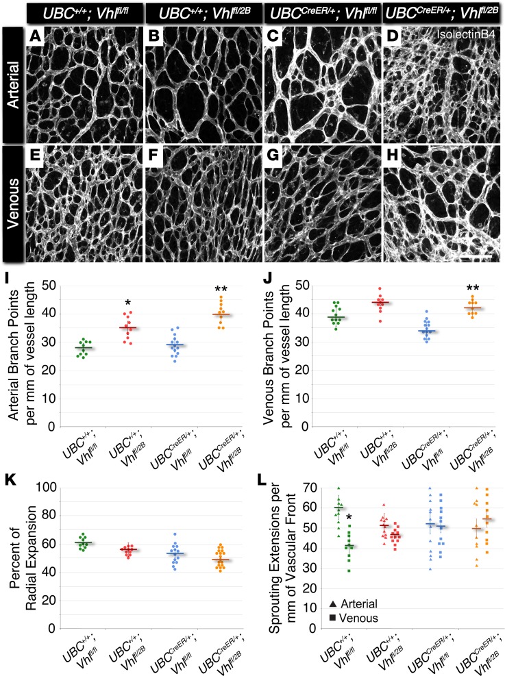
Figure 1. The Vhl 2B mutation causes increased arterial and venous branching in P5 mouse retinas without changes in vessel expansion or endothelial cell sprouting.
(A–H) Representative images of P5 mouse retinal vasculature stained with isolectin B4. Arterial (A–D) and venous (E–H) regions are shown for UBC+/+Vhlfl/fl, UBC+/+Vhlfl/2B, UBCCreER/+Vhlfl/fl, and UBCCreER/+Vhlfl/2B littermates exposed to tamoxifen. Scale bar: 100 μm. (I) Arterial branch points per vessel length measured from P5 vascular networks. *P ≤ 0.05 vs. UBC+/+Vhlfl/fl. **P ≤ 0.01 vs. UBC+/+Vhlfl/fl and UBCCreER/+Vhlfl/fl. (J) Venous branch points per vessel length measured from P5 vascular networks. **P ≤ 0.01 vs. UBCCreER/+Vhlfl/fl. (K) Percent of radial expansion of retinal vessels from the optic disc toward the peripheral edge. No significant differences found. (L) Endothelial sprouting extensions per vessel length of the vascular front for arterial (triangles) and venous (squares) regions. *P ≤ 0.05 vs. UBC+/+Vhlfl/fl arterial. All others are not significantly different. Values are averages ± SEM. n = 10–15 retina images per group (i.e., 2–3 from 5 experimental litters). All statistical comparisons performed using 1-way ANOVA followed by pair-wise comparisons with a 2-tailed Student t test.

