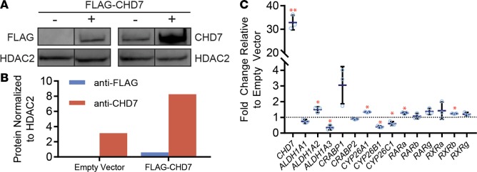
Figure 3. Increased CHD7 affects expression of retinoic acid–related genes.
(A and B) 293T cells transfected with a vector containing human CHD7 and FLAG. CHD7 and FLAG levels were assayed by Western blot (A) and quantified (B) using ImageJ software. This experiment was repeated twice. Images were derived from duplicate samples run contemporaneously on parallel gels. (C) Expression of retinoic acid–related genes examined using qPCR of mRNA extracted from 293T cells transfected with CHD7 overexpression or empty vector. This experiment was repeated 2 times. Significance was determined by Student’s t tests. *P ≤ 0.05.

