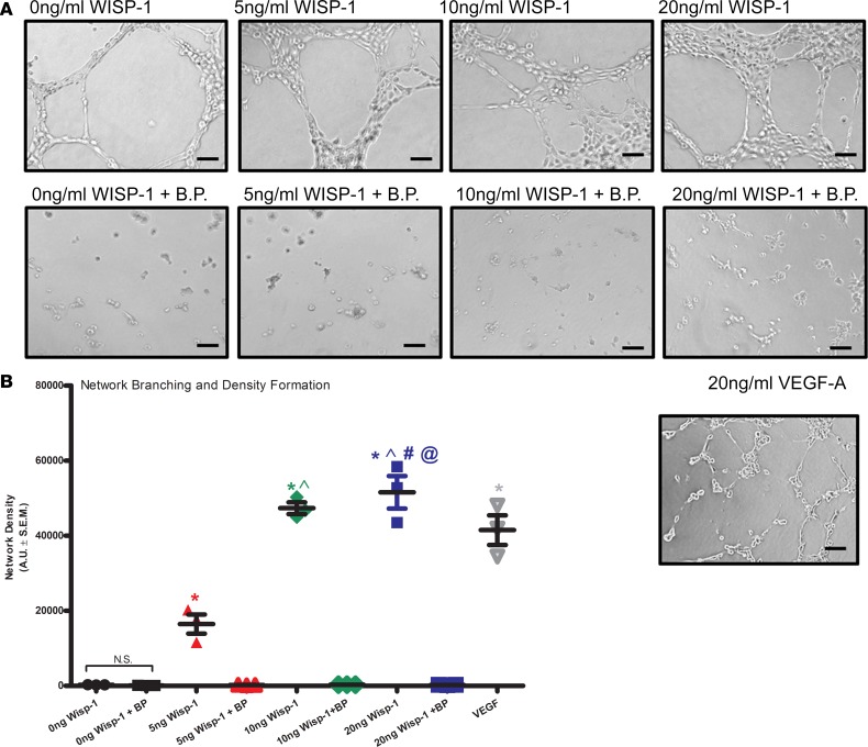
Figure 11. WISP-1 enhances endothelial cell branching and network densities in vitro.
(A) Thirty thousand HCAECs were seeded in triplicate in 48-well plates on growth factor–reduced Matrigel in the presence of 0 ng/ml (vehicle), 5 ng/ml, 10 ng/ml, or 20 ng/ml recombinant WISP-1 and or BP, or 20 ng/ml VEGF-A. Scale bars: 100 μm. (B) Graph represents density of network branching determined by software analyses of images (5/well). A is representative of 4 repeated analyses. B was performed in triplicate of each experimental groups and are representative of 3 independent experiments. Results depicted as mean ± SEM,*P ≤ 0.05 relative to control, ^P ≤ 0.05 relative to 5 ng/ml, #P ≤ 0.05 relative to 10 ng/ml, and @P ≤ 0.05 relative to VEGF-A (10ng/ml). P values obtained by 1-way with Bonferroni’s post test.

