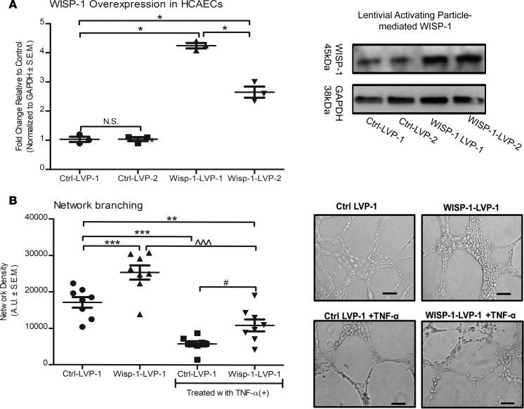
Figure 14. Constitutive overexpression of WISP-1 functionally enhances endothelial cell network density.
(A) HCAECs were transduced with either control, nontargeting lentiviral activation particles (Ctrl-LVP), or WISP-1 lentiviral activation particles (WISP-1-LVP) for constitutive activation. (B) Clone, WISP-1-1 (most robust activation relative to control) was used to assess network density abilities. Thirty thousand cells from each experimental group were seeded in triplicate in 48-well plates on growth factor–reduced Matrigel. In parallel, cells were also treated with TNF-α (10 ng/ml). Density of network branching was determined by software analyses of images (5/well) after 8 hours. Graphs show branching density determined by software analyses. Analysis of Western blots are from the same experimental group, repeated 3 times. Data in B are from experiments performed in triplicate; n = 8 per group (average density of 5 photographs per group). Results depicted as mean ± SEM of AUs *P ≤ 0.05, **P ≤ 0.01, ***P ≤ 0.001, relative to Ctrl–LVP-1; ^^^P ≤ 0.001, relative to Wisp-1–LVP-1; #P ≤ 0.001, relative to Ctrl–LVP-1 with TNF-α. P values obtained by 1-way ANOVA with Bonferroni’s post test.

