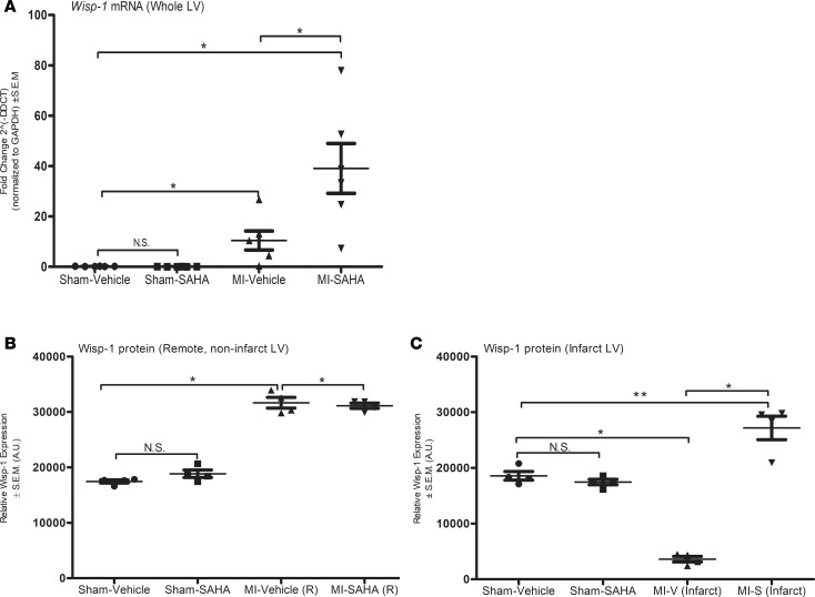
Figure 3. Wisp-1 mRNA and Wisp-1 protein expression are increased in response to MI injury and HDAC inhibition within the LV 7 days after MI.
(A) qPCR of Wisp-1 expression from LV at 7 days after MI. Data represents the 2–ΔΔct and are normalized to Gapdh CT values. Western blot analysis of Wisp-1 protein in (B) remote noninfarct LV and (C) infarcted LV of post-MI myocardium or sham control tissue. Data are normalized to Gapdh. A is from 6 mice per group. qPCR was performed in triplicated and repeated 3 times. B and C are from 4 mice per group and representative of 3 independent studies. Results depicted as mean ± SEM. *P ≤ 0.05, **P ≤ 0.01. P values obtained by 1-way ANOVA with Tukey’s post test.

