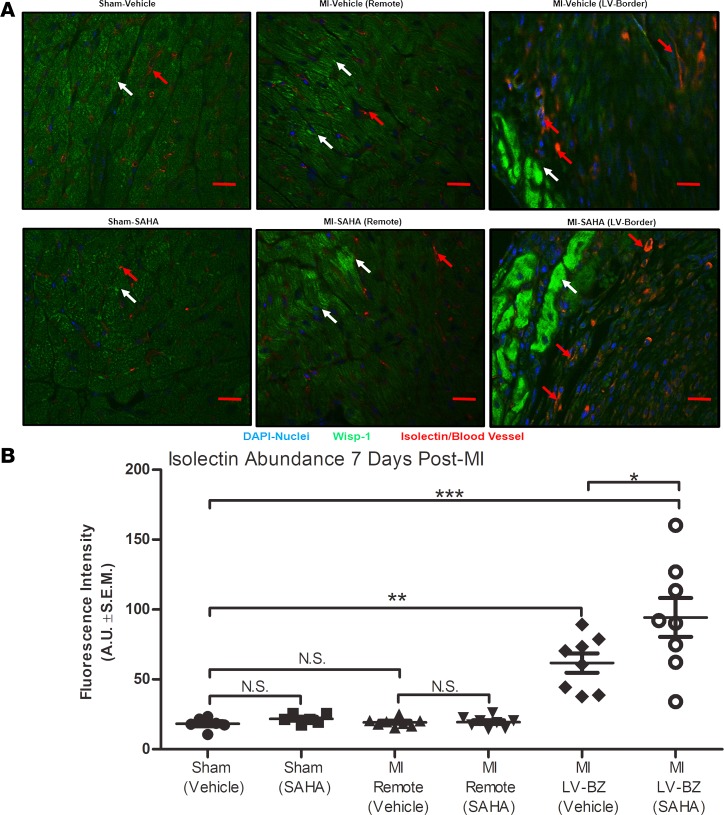
Figure 5. Wisp-1 expression is proximal to enhanced micro-vessel density at the border zone 7 days after MI.
Ten- to 12-week-old male CD1 mice were subjected to either sham (control) or ligation of the coronary artery surgery and received daily i.p. injections of DMSO (vehicle-control) or HDAC inhibition, SAHA (25 mg/kg). Seven days after MI, mice were euthanized. (A) Localization and abundance of Wisp-1 (white arrows) and microvasculature (red arrows), 10× magnification, scale bars: 20 μm. (B) Relative fluorescence intensity (green/FITC) per field was determined using Image J (Fiji), normalized to nuclei (DAPI/blue) and quantified by AUs. A and B are from 6 mice/sham and 8 mice/MI group and are representative of 2 independent studies. Results depicted as mean ± SEM, *P ≤ 0.05, **P ≤ 0.01, ***P ≤ 0.00. P values obtained by 1-way ANOVA with Tukey’s post test.

