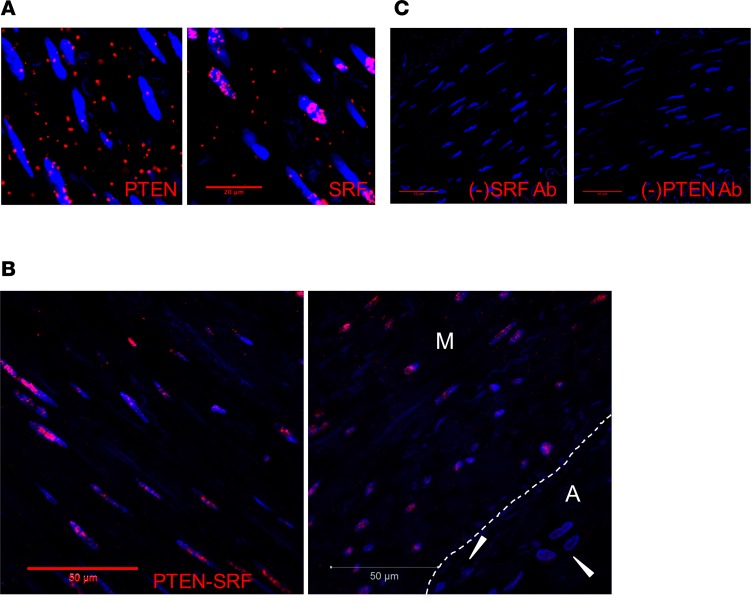
Figure 1. Interaction of PTEN and serum response factor (SRF) in the nucleus of medial smooth muscle cells (SMCs).
Proximity ligation assay (PLA) and confocal microscopy were used to detect PTEN-SRF interactions in medial SMCs of human nonatherosclerotic coronary arteries. Representative images shown. (A) Left: PTEN positive control using mouse anti-PTEN primary antibody and anti-mouse PLUS and anti-mouse MINUS PLA probe demonstrates nuclear and cytoplasmic expression of PTEN. Right: SRF positive control using rabbit anti-SRF primary antibody and anti-rabbit PLUS and anti-rabbit MINUS PLA probe demonstrates predominantly nuclear expression of SRF. Scale bar: 20 μm. (B) PLA using mouse anti-PTEN and rabbit anti-SRF primary antibodies and anti-mouse PLUS and anti-rabbit MINUS PLA probe demonstrates PTEN-SRF nuclear interactions in medial SMCs of human nonatherosclerotic hyperplasia coronary arteries, but no interactions in adventitial cells (arrowheads; right panel). M = media; A = adventitia; white dashed line in right panel indicates the external elastic lamina. Scale bars: 50 μm. (C) PLA negative controls for SRF (left) and PTEN (right) demonstrate lack of signal when either primary antibodies are omitted. Scale bars: 50 μm. For all panels: Red = positive PLA; Blue = DAPI for cell nuclei.

