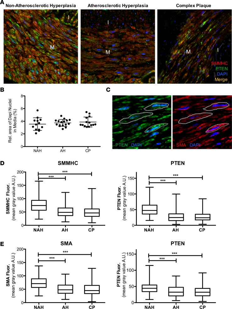
Figure 3. Single-cell analysis of PTEN and smooth muscle myosin heavy chain (SMMHC) α-smooth muscle actin (αSMA) expression in medial SMCs.
(A) Representative PTEN (green) and SMMHC (red) stained confocal images of lesions representing nonatherosclerotic hyperplasia (NAH), atherosclerotic hyperplasia (AH; intima > 200 μm), or complex plaque (CP); merged images shown. M = arterial media; I = arterial intima. (B) Relative area of DAPI expression in the media per vessel was measured by ImageJ 1.47v as a measure of cellularity and averaged from 4 or 5 confocal images (original magnification, ×63) of media per vessel. NAH: N = 13 individual vessels from 7 independent hearts; AH: N = 16 individual vessels from 10 independent hearts; CP: N = 14 individual vessels from 11 independent hearts. (C) Representative confocal images of arterial media from NAH vessels immunofluorescently stained for PTEN (green; left) and αSMA (red; right); nuclei are stained with DAPI (blue). White dashed lines represent cell boundaries for ROI measurements. PTEN and SMMHC/αSMA levels within cell boundary ROI were measured by ImageJ 1.47v as described in Methods. (D) The mean gray value within cell boundary ROI for single-cell analysis of medial smooth muscle cells (SMCs) for SMMHC (left) and PTEN (right) levels was determined using ImageJ. NAH: N = 361 individual cells from 8 vessels and 5 independent hearts; AH: N = 294 individual cells from 7 vessels and 6 independent hearts; CP: N = 286 individual cells from 6 vessels and 5 independent hearts. ***P ≤ 0.001. Plotted data include the median gray value (horizontal bar), interquartile range (box boundary) and minimum to maximum range of data points (vertical bar). (E) The mean gray value within cell boundary ROI for single-cell analysis of medial SMCs for αSMA (left) and PTEN (right) levels was determined using ImageJ. NAH: N = 196 individual cells from 6 vessels and 6 independent hearts; AH: N = 197 individual cells from 6 vessels and 5 independent hearts; CP: N = 195 individual cells from 6 vessels and 5 independent hearts. ***P ≤ 0.001 by Kruskal Wallis with Dunn’s posttest comparisons.

