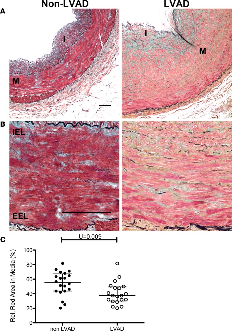
Figure 5. Smooth muscle dedifferentiation in coronary arteries exposed to CF-LVAD.
Coronary arteries from explanted hearts of non– left ventricular assist device (non-LVAD) or continuous-flow LVAD (CF-LVAD) patients were stained with Movat’s pentachrome stain. (A and B) Representative low-power (A) and high-power (B) stained images showing decreased red staining in medial and intimal smooth muscle cells in CF-LVAD–exposed (right) compared with non–LVAD-exposed (left) vessels, indicative of decreased muscle. M = arterial media; I = arterial intima; IEL = internal elastic lamina; EEL = external elastic lamina.Scale bars: 100 μm. (C) The percentage red-stained area of the media was measured by ImageJ and averaged from 4 or 5 confocal images (original magnification, ×40) of media per vessel. Non-LVAD: N = 20 individual vessels from 11 independent hearts; CF-LVAD: N = 22 individual vessels from 12 independent hearts. **P = 0.009 by Mann-Whitney 2-tailed U test. Horizontal lines show the median and interquartile range.

