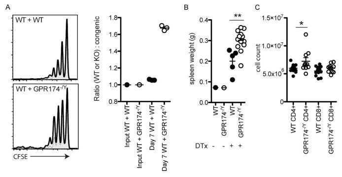
Figure 1.
GPR174 constrains T cell proliferation in vivo. (a) To assess homeostatic proliferation, a 1:1 mixture of wild-type (CD45.1/2+; shaded gray plots) and either wild-type (CD45.2+; upper left panel; open solid line) or Gpr174−/Y (CD45.2+; lower left panel; open solid line) naive CD8+ T cells were labeled with CFSE. A total of 1×10⁶ T cells were transferred into CD45.1+ recipient mice that had been irradiated the day before with 600 cGy irradiation. Homeostatic proliferation was assessed based on the CFSE dilution profile (left) and ratio of wild-type to wild-type or Gpr174−/Y cells recovered in the spleen of recipient mice seven days after transfer was determined (right). (b–c) To assess endogenous T cell proliferation in a Treg cell ablation model, cohorts of wild-type and Gpr174−/Y mice that expressed a Foxp3-DTR transgene were treated with diphtheria toxin intrapetironeally with an initial dose of 20 μg kg−1 on day 0, and then with 5 μg kg−1 on days 2 and 4. Spleen weights (b) and numbers of CD4+ and CD8+ T cells in the spleen (c) were quantified on day 7; * P < 0.05; ** P < 0.01. Data are representative of two independent experiments; each dot represents a mouse.

