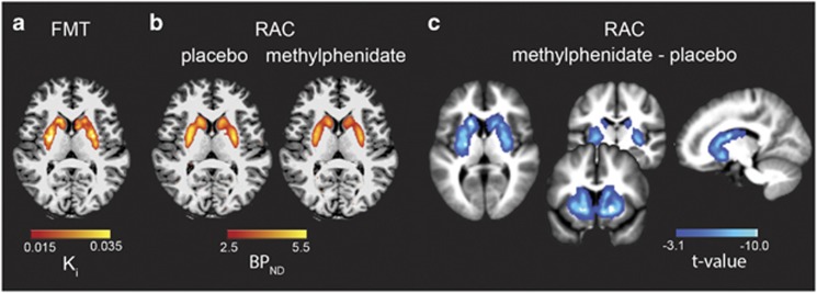
Figure 1.
Within-subject measures of dopamine synthesis capacity and D2/3 receptor binding. (a) [18F]FMT Ki signal reflecting dopamine synthesis capacity was measured throughout striatum. The axial slice illustrates the extent of the striatal Ki signal for a representative subject overlaid on the subject’s native space T1 MPRAGE. (b) [11C]raclopride BPND displayed for placebo (baseline) scan as well as post-methylphenidate scan for the same representative subject. Methylphenidate administration reduced [11C]raclopride BPND. (c) Striatal regions showing significantly reduced [11C]raclopride BPND following methylphenidate administration across all participants. The t-map for the paired t-test comparing baseline and post-methylphenidate [11C]raclopride BPND is displayed on the normalized mean T1 MPRAGE for all subjects.

