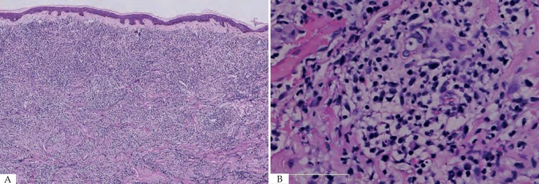
Figure 2.
A - Diffuse dermal atypical myeloid infiltrates involving subcutis with Grenz zone. (Hematoxylin & eosin, x10); B - Infiltrates were composed of small to medium sized mononuclear cells with atypical cytological features, including nuclear hyperchromasia, coarse chromatin pattern, and thickened nuclear membrane. (Hematoxylin & eosin, X40)

