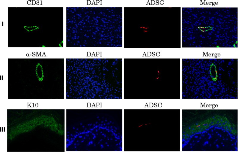
FIGURE 4.

Expression of epithelial cell markers CD31, α-SMA, K10 in wound skin. (I) Photomicrographs showing Endothelial cells immunostaining for CD31 (green), engrafted ADSCs (red) in the wound site of diabetic rats, nuclear counterstained with DAPI (blue) at 200× magnification. (II) Images showing vascular smooth muscle cells immunostaining for α-SMA (green), engrafted ADSCs (red) and DAPI (blue) in the wound site of diabetic rats. (III) Images showing epidermal epithelium immunostaining for K10 (green), engrafted ADSCs (red) and DAPI (blue) in the wound site of diabetic rats. Scale bar = 50 μm
