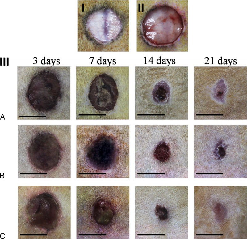
FIGURE 6.

Macroscopic lesion appearance. Diabetic rats model of skin injury were used (I,III). A standardized wound area (1.5-cm-diameter circle skin in full thickness removed) was induced on the dorsal surface of diabetic rats (I) and normal rats (II) (they have similar weight and age). Compared with normal rats, diabetic rats have poor blood supply, lack of subcutaneous tissue, thin skin and poor elasticity. (III) Lesions appearance at 3, 7, 14 and 21 days.
