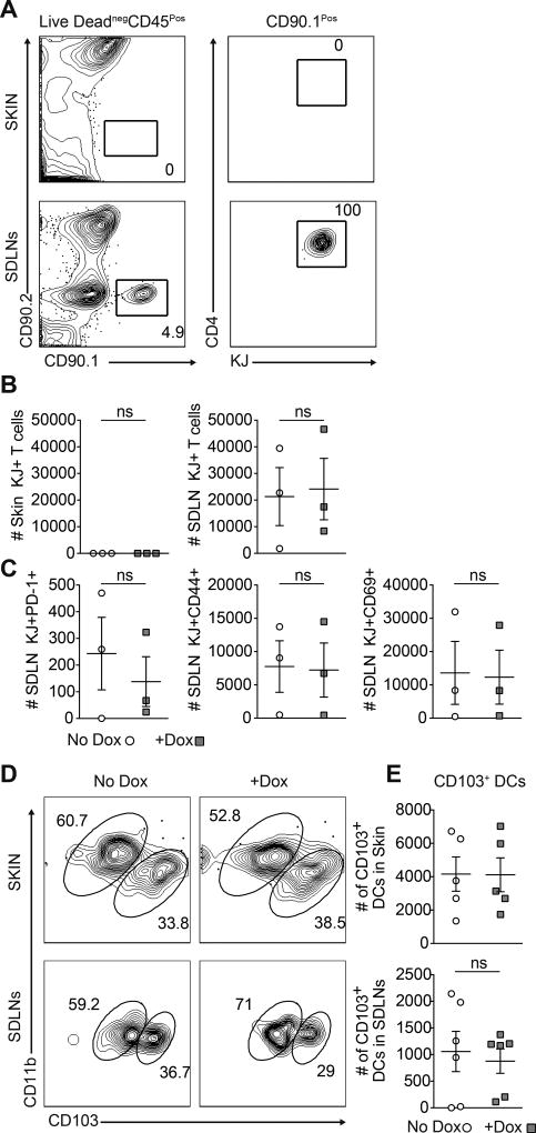
Figure 5. The presence of antigen-specific T cells in SDLNs is insufficient to drive CD103+ DDCs emigration from skin.
(A) Representative flow cytometric plots and (B) quantification of adoptively transferred DO11 cells in the skin and SDLNs of No Dox controls and Dox treated K5/TGO/TCRα−/− mice on day 2. (C) Quantification of activation marker expressing DO11 cells in the SDLNs. (D) Representative flow cytometric plots and (E) quantification of CD103+ DDCs in the skin and SDLNs of No Dox controls and Dox treated K5/TGO/ TCRα−/− mice on day 2. One representative experiment of three is shown. ns = no significant difference. Data are mean ± s.e.m, Student’s unpaired t-test (B. C, E).

