. 2018 Mar 28;7:e32579. doi: 10.7554/eLife.32579

Figure 1. Construction of codon altered JFH1.

(A) Cell cultures coinfected with two strains of HCV result in four populations: uninfected, two types of singly infected, and coinfected cells. (B) Three segments of the JFH1 genome, that were roughly 1000 nucleotides in length and had altered codon usage, were designed using GeneArt algorithms and synthesized. These genome fragments were then cloned into the JFH1 strain of HCV. Huh7.5.1 cells were transfected with each construct using electroporation to create long-term HCVcc cultures and viability was followed over time by immunoblotting of cell lysates for the HCV core protein. (C) Viral passages shown in (B) were monitored by qRT-PCR analysis of viral RNA in culture supernatants. Only the CA-3 virus demonstrated growth kinetics comparable to wild-type (WT) JFH1.
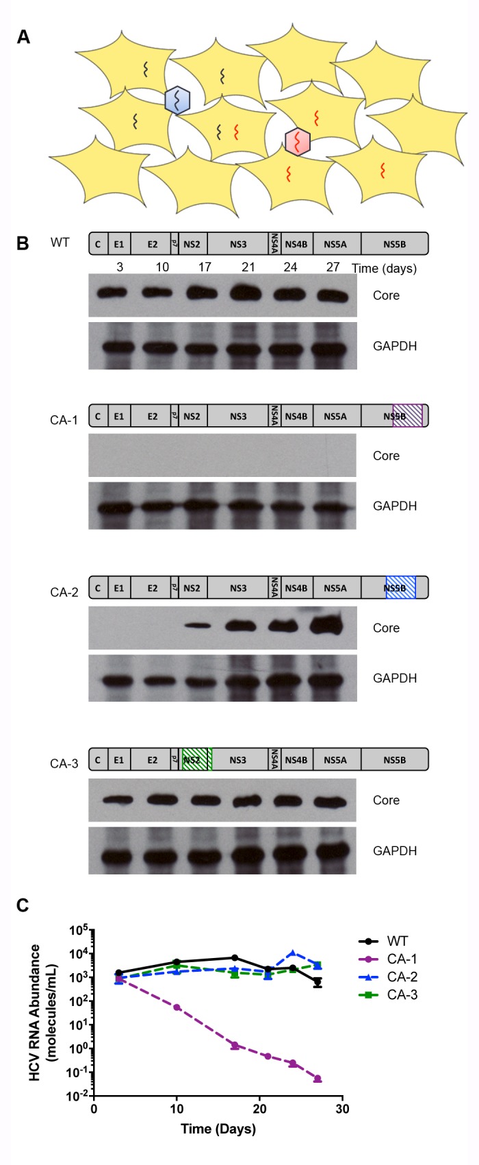
Figure 1.

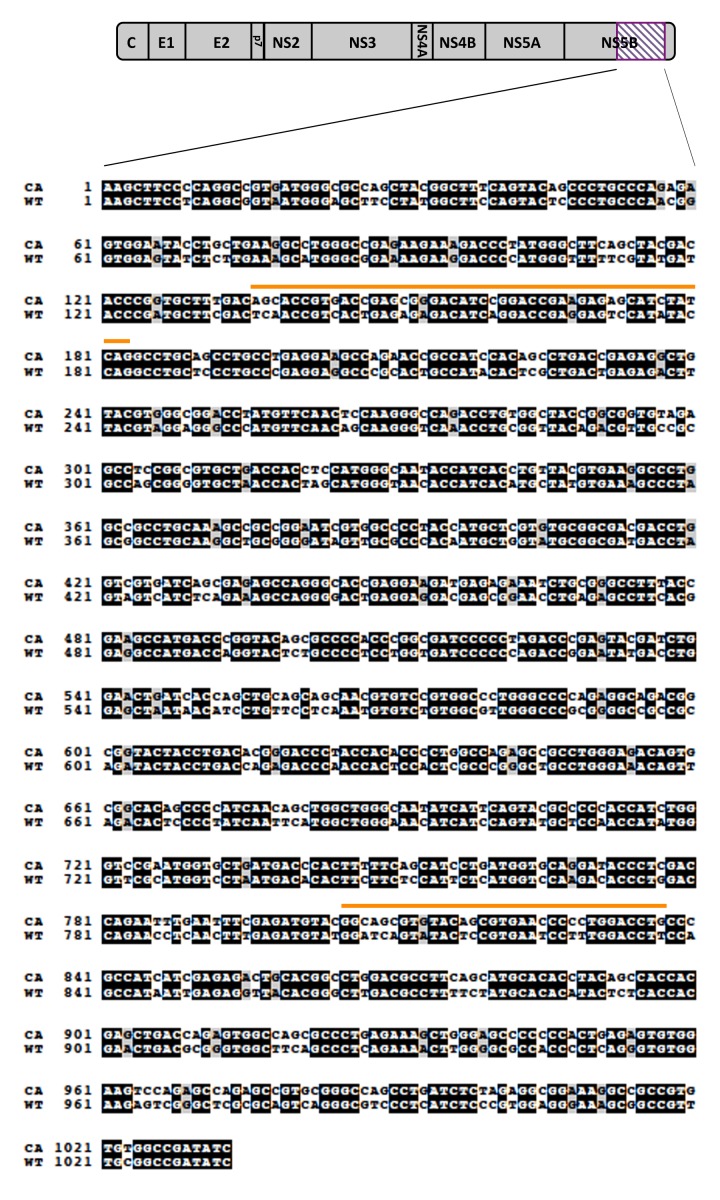
Figure 1—figure supplement 1. Alignment of codon-altered and wild-type JFH1 viral RNAs.

Figure 1—figure supplement 1.

Sequence alignments were generated using ClustalW to highlight the nucleotide changes incorporated into the CA-1 viral RNA. Sequences highlighted with orange bars identify regions containing covarying mutations indicating functional RNA secondary structures.
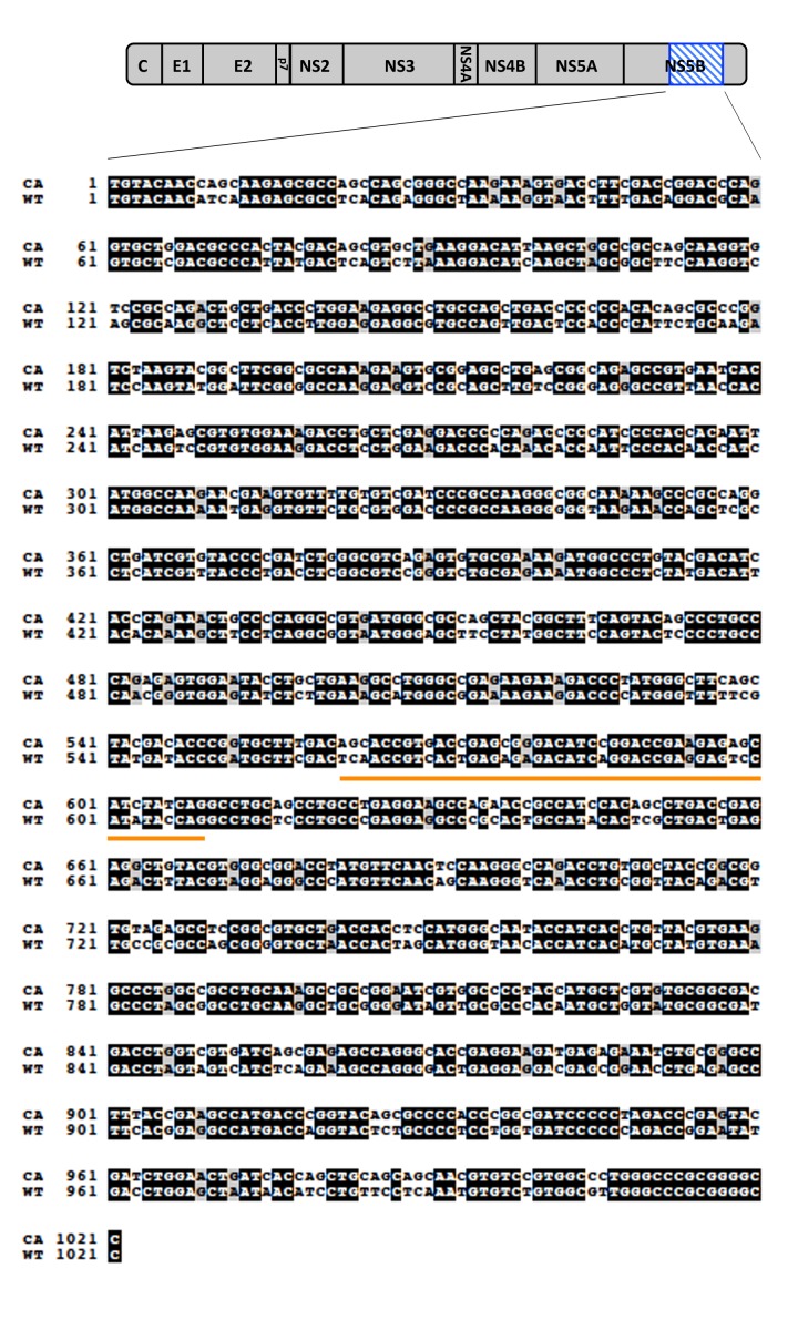
Figure 1—figure supplement 2. Alignment of codon-altered and wild-type JFH1 viral RNAs.

Figure 1—figure supplement 2.

Sequence alignments were generated using ClustalW to highlight the nucleotide changes incorporated into the CA-2 viral RNA. Sequences highlighted with orange bars identify regions containing covarying mutations indicating functional RNA secondary structures.
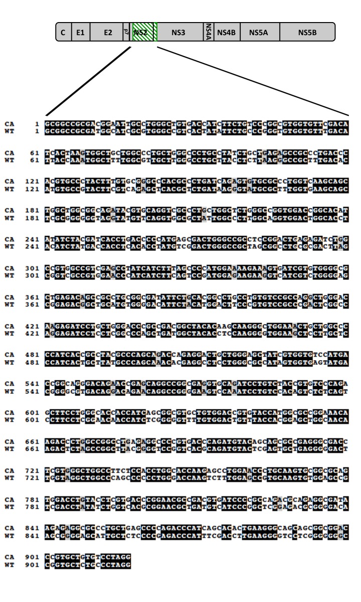
Figure 1—figure supplement 3. Alignment of codon-altered and wild-type JFH1 viral RNAs.

Figure 1—figure supplement 3.

Sequence alignments were generated using ClustalW to highlight the nucleotide changes incorporated into the CA-3 viral RNA. No regions of covarying mutations were observed within this region.