. 2018 Mar 28;7:e32579. doi: 10.7554/eLife.32579

Figure 3. Flow cytometry to test dominance of viruses resistant to NS3 protease inhibitor.

(A) Structure of protease inhibitor BILN-2061. (B) Structure of NS3 protein. D168 (red) is located in the protease domain adjacent to the active site (lavender). D168A confers resistance to BILN-2061. (C) Diagram of CA virus with altered sequence (green hatches) and WT virus with location of D168A mutation identified. (D) The four types of cells present in the absence of inhibitors are uninfected (U), infected with drug-susceptible virus cells (S), infected with both drug-susceptible and drug-resistant virus (S + R) and drug-resistant virus (R). In the presence of a DAA, three outcomes are possible and are indicated by the changing density of the cell populations: (E) Drug-resistance is cis-dominant, (F) Drug resistance is dominant and (G) Drug susceptibility is dominant. Huh7.5.1 cells were coinfected with CA and WT-D168A for 72 hr followed by treatment with (H/I) DMSO or (J/K) 2 μM BILN-2061 for 36 hr. Cells were stained with CA and WT vRNA probes and analyzed by flow cytometry (H, J) and results from three replicates quantified (I, K). NS3 drug-resistance was found to be cis-dominant.

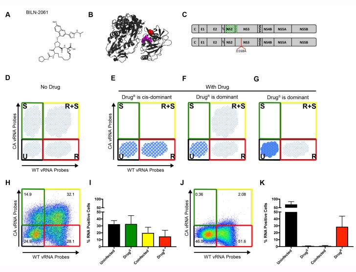
Figure 3.

Figure 3—figure supplement 1. NS3-D168A confers resistance to BILN-2061 in both the wild-type and the codon-altered backgrounds.

Figure 3—figure supplement 1.

D168A was cloned into both the WT (left) and CA (right) backgrounds and tested for resistance to BILN-2061. Huh7.5.1 cells were infected with WT, WT-D168A, CA or CA-D168A viruses at MOI = 0.1 FFU/cell in the absence or presence of 2 μM BILN-2061 as indicated. Viral RNA was harvested from culture supernatants collected at 72 hr post infection and analyzed by qRT-PCR.
Figure 3—figure supplement 2. Exogenously expressed drug-resistant NS3 and NS5A do not rescue drug-susceptible HCV in trans.

Figure 3—figure supplement 2.

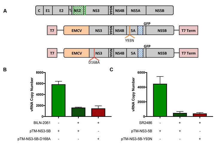
(A) Schematic of codon-altered HCV RNA, and non-structural protein expression plasmids (pTM-NS3-5B) that contain either no mutation, the D168A mutation that confers resistance to BILN-2061 or the Y93N mutation that confers resistance to SR2486. (B) Huh7-Lunet-T7 cells were infected with CA virus for 72 hr and then transfected with wild-type or D168A pTM constructs in the absence and presence of 2 μM BILN-2061 for 24 hr as indicated. Viral RNA was harvested from cell culture supernatants and quantified by qRT-PCR. Results from samples harvested in triplicate are shown. (C) Cells were infected with CA virus for 72 hr and then transfected with wild-type or Y93N pTM constructs in the absence and presence of SR2486 for 24 hr as indicated. Viral RNA was harvested from cell culture supernatants and quantified by qRT-PCR. Results from samples harvested in triplicate are shown.
Figure 3—figure supplement 3. Drug resistance to R1479 confers major fitness cost to JFH1.

Figure 3—figure supplement 3.

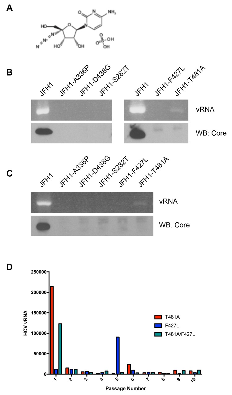
(A) R1479 is a nucleotide analog inhibitor of the HCV NS5B polymerase. (B) WT virus was passaged every 72 hr ten times in the presence of 25 μM R1479. Extracted vRNA from each passage was quantified and sequenced to determine if selection of resistance associated variants occurred. Of the variants identified by sequencing only T481A and F427L demonstrated any replication capacity as measured by core expression in transfected cells or the presence of vRNA in culture supernatants at day (B) 7 or (C) 21 post transfection. (D) We attempted to select for compensatory mutations that increased the fitness of WT-T481A, WT-F427L or the double mutant WT-T481A/F427L by passaging these viruses in the presence of 25 μM R1479 ten times and sequencing total viral RNA from these passages. No compensatory mutations were identified.