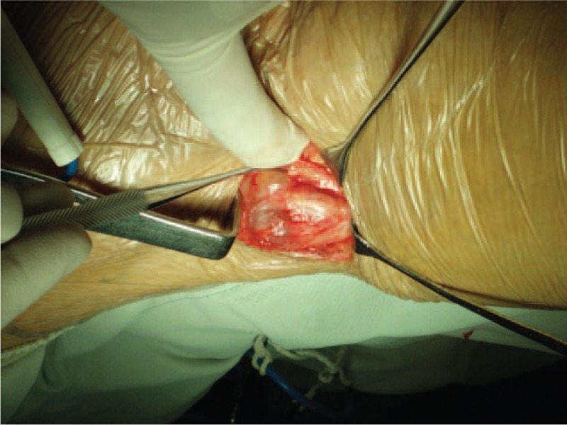
Figure 2.

Intraoperative photograph of the popliteal fossa. A popliteal cyst (marked by the long arrow) sized about 3×2×3 cm3 extending from the proximal to the distal end of the popliteal fossa. It tightly encased left common peroneal nerve (marked by the short arrow) at the level of 8 cm away from capitulum fibulae.
