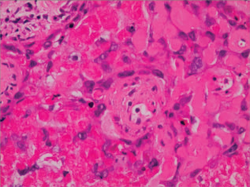
Figure 3.

High-power photomicrograph (B, hematoxylin–eosin, original magnification ×200) showed epithelioid cells with necrotic debris and peritumoral hyaline-like material.

High-power photomicrograph (B, hematoxylin–eosin, original magnification ×200) showed epithelioid cells with necrotic debris and peritumoral hyaline-like material.